A better approach to assess liver viability during organ donor evaluation
Ernesto Duarte Tagles1,2,3, Luis Carlos Rodríguez Sancho3, Jorge Rubén Béjar Cornejo4,5, Mario Alberto Flores León1,3, Alejandro Lugo Baruqui3, Salvador Castillo Barón3, Marisela Correa Valdéz3, Aarón David Luna Espinoza4,5, Martha Susana Pérez Cornejo2,3, José Armando Portugal Lazcano3.
1Department of transplantation, Hospital General del Estado "Dr. Ernesto Ramos Bours", Hermosillo, , Mexico; 2Organ Donation Coordination, Hospital General del Estado "Dr. Ernesto Ramos Bours", Hermosillo, , Mexico; 3Department of transplantation, Hospital CIMA Hermosillo, Hermosillo, , Mexico; 4Department of radiology, Unidad Diagnóstica Avanzada, Hermosillo, , Mexico; 5Deparment of Radiology, Hospital General del Estado "Dr. Ernesto Ramos Bours", Hermosillo, , Mexico
Introduction: The lack of livers for transplant is a worldwide reality and in Mexico this condition might be even worse. For May 2023 in Mexico there were 206 patients waiting for a liver transplant. In 2022 we performed 238 liver transplants out of 443 brain death donors (none of them were non heart beating donors). Reasons why 46.2% of livers are not use are wide but two are among the principals: 1) A high prevalence of overweight and obesity in mexican population, meaning a high frecuency of liver steatosis and 2) a poor air connectivity among mexican cities, increasing the risk of organ failure due to prolonged ischemia time. Because the common study to evaluate liver steatosis is ultrasound despite a low sensitivity, we proposed a better way to assess liver conditioin before harvesting, diminishing money spent and assuring better transplant results.
Method: A prospective double blinf study was conducted in Hospital General del Estado from October 2015 to November 2021, consisting in that in every donor, no matter body mass index, a liver ultrasound was made by a radiologist and latter as well as an elastography (using a FIbroscan 502 model) by a second radiologist in different times. Then results were confronted during organ harvesting by surgeon expertise sight evaluation, who decided to take the liver or not.
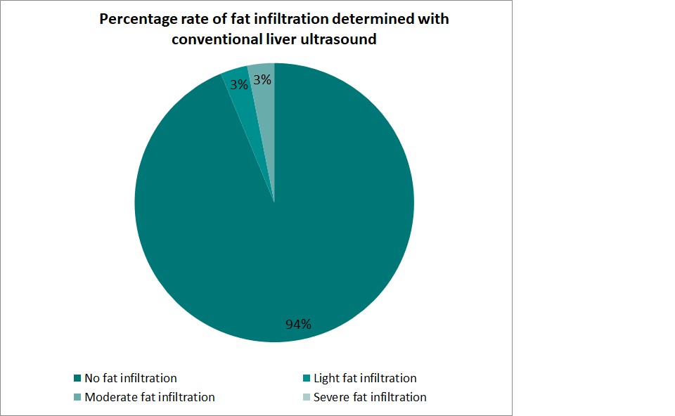
Results: We had 32 brain death organ donors to whom an ultrasound and an elastography was made. Median age was 41 years and weight was 74.66 kgs, with a range from 55 to 120 kgs. Out of the 32 donors, 93.8% were diagnosed without liver steatosis, 3.1% with mild and 3.1% with severe steatosis with ultrasound:

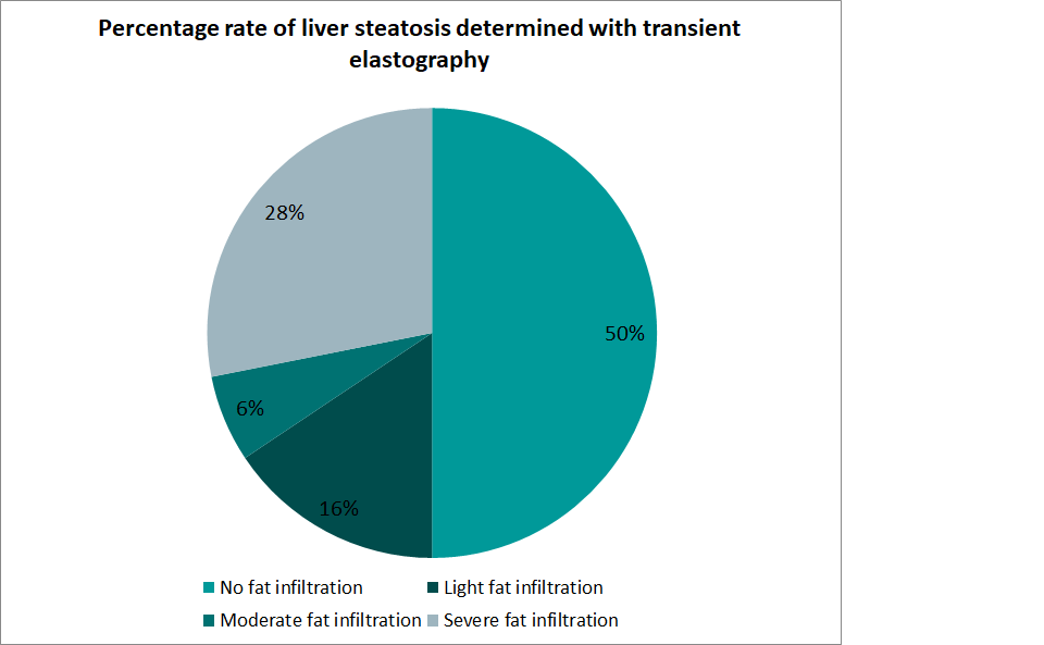
while elastography showed 50% no steatosis, 15.6% light, 6.3% mild and 28.1% severe steatosis.

Conclusion: Our study showed that using ultrasound to evaluate liver condition for using in transplantation regarding fatty infiltration is not as good as it might be thought. For no liver steatosis, concordance between ultrasound and elastography was 50% but in cases for severe fatty liver infiltration, diagnosed rate was 1 Vs 9 for ultrasound Vs elastography, this means that for every case of severe steatosis diagnosed by ultrasound, 9 were identified with elastography and these results were confirmed at the moment of surgery. This shows that elastography is much better in identifing mild/severe steatosis in organ donors than ultrasound and thus becoming a better resource in the evaluation of the liver for using for transplantation in brain death organ donor, leading to a better control of money spent but most important, in preventing a primary organ failure after transplantation.